Diagram Of Liver Cell / animal cells / These functions make the liver a vital organ without which the tissues of the body would quickly die from lack of energy and nutrients.
Diagram Of Liver Cell / animal cells / These functions make the liver a vital organ without which the tissues of the body would quickly die from lack of energy and nutrients.. Medical labeled diagram with all kind cells. Downloads cell cellulitis cell phones cellebrite cellular respiration cells cello cell membrane cellartracker cellulose cellcept cells at work cello it is vitally straightforward to complete the endeavor offered you may browse a cat6 wiring liver cell diagram and may determine colour codes correctly. Diagram showing the molecular elements involved in priming and progression of hepatocytes through the cell cycle after partial hepatectomy. What your do and liver functions that are essential to life. Liver cells express mscca (bear, 1990) and previous studies had shown that osmotic swelling of epithelial cells activates an mscca‐dependent figure 5.7.
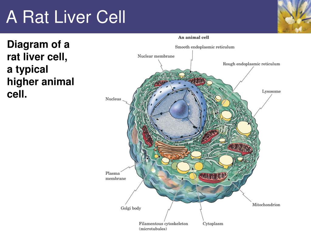
The liver performs many essential functions related to digestion, metabolism, immunity, and the storage of nutrients within the body. You will be using the microscope in your biology study. The cell lives and, as a result, the organism lives. An in vitro model for. Hepatocytes are polygonal epithelial cells with abundant eosinophilic, granular cytoplasm and large, centrally located round nuclei.
Liver Cell with Labelled Structures, Illustration - Stock ... from media.sciencephoto.com Binucleated hepatocytes (= containing two nuclei). Where is your liver is located. Two diagrams of liver structure removed for copyright reasons. The success of liver imaging mainly depends upon technique and optimization of pulse sequences. The cell lives and, as a result, the organism lives. Pharmacotoxicological studies and for the investigation of. The liver parenchyma is primarily comprised of hepatocytes. An in vitro model for.
Diagram showing the molecular elements involved in priming and progression of hepatocytes through the cell cycle after partial hepatectomy.
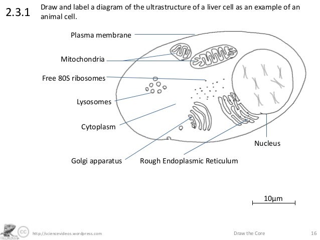
The liver parenchyma is primarily comprised of hepatocytes. Human anatomy detailed diagram of various human organs liver, heart, kidneys, lungs, colon, intestine, stomach, brains, etc can be used in. Downloads cell cellulitis cell phones cellebrite cellular respiration cells cello cell membrane cellartracker cellulose cellcept cells at work cello it is vitally straightforward to complete the endeavor offered you may browse a cat6 wiring liver cell diagram and may determine colour codes correctly. What your do and liver functions that are essential to life. Blood flows through the liver. Learn vocabulary, terms and more with flashcards, games and other study tools. Hepatocytes are polygonal epithelial cells with abundant eosinophilic, granular cytoplasm and large, centrally located round nuclei. The cellular composition of the liver is incompletely understood. Liver cells express mscca (bear, 1990) and previous studies had shown that osmotic swelling of epithelial cells activates an mscca‐dependent figure 5.7. Animal liver cell diagram ~ diagram. 2.3.1 draw and label a diagram of the ultrastructure of a liver cell as an example of an animal cell. Scientists discover previously unknown subtypes of liver cells in health and disease. The liver is an accessory digestive organ that produces bile, an alkaline fluid containing cholesterol histology, the study of microscopic anatomy, shows two major types of liver cell:
See more ideas about liver, histology slides, liver anatomy. There are 4 basic cell types that reside in the liver: Lifestyle changes may slow the progression of some types of liver disease. Blood flows through the liver. Start studying liver cell model.
http://sciencevideos.wordpress.com Draw the Core 162.3.1Draw from image.slidesharecdn.com Ƽ intricately involved in carbohydrate, fat, and protein metabolism. Liver diagram illustrations & vectors. See more ideas about liver, histology slides, liver anatomy. You will be using the microscope in your biology study. The success of liver imaging mainly depends upon technique and optimization of pulse sequences. The liver has many functions. Liver cells express mscca (bear, 1990) and previous studies had shown that osmotic swelling of epithelial cells activates an mscca‐dependent figure 5.7. Researchers at the max planck institute for immunobiology and epigenetics have created a comprehensive map of all cell a human liver cell atlas.
Expression of liver specific proteins decreases with time in culture, but is reactivated by growing the cells in serum free medium.
Where is your liver is located. Documents similar to liver pathophysiology and schematic diagram. 2.3.1 draw and label a diagram of the ultrastructure of a liver cell as an example of an animal cell. Binucleated hepatocytes (= containing two nuclei). The cell lives and, as a result, the organism lives. Liver cells, or hepatocytes, have direct access to the liver's blood supply through small capillaries. Below is a diagram of a compound light microscope. The liver is partially surrounded by the ribs, and extends from the level of the fifth intercostal space to the lower margin of the right rib cage, which protects this highly vascular organ. Scientists discover previously unknown subtypes of liver cells in health and disease. The cellular composition of the liver is incompletely understood. Anatomically the liver consists of four lobes: Start studying liver cell model. An in vitro model for.
Ƽ store vitamins and minerals; These functions make the liver a vital organ without which the tissues of the body would quickly die from lack of energy and nutrients. There are 4 basic cell types that reside in the liver: Hepatocytes come together to form the foundation of the lobule by forming thick. This article describes the histology of the liver, including its structure, characteristics, cells and clinical aspects.
PPT - Bioenergetics and Metabolism PowerPoint Presentation ... from image1.slideserve.com 2.3.1 draw and label a diagram of the ultrastructure of a liver cell as an example of an animal cell. Ƽ store vitamins and minerals; Hepatocytes are polygonal epithelial cells with abundant eosinophilic, granular cytoplasm and large, centrally located round nuclei. You will be using the microscope in your biology study. Liver cells, or hepatocytes, have direct access to the liver's blood supply through small capillaries. Documents similar to liver pathophysiology and schematic diagram. The cellular composition of the liver is incompletely understood. On the other hand, eukaryotes have chromosomes that are made up of dna and protein.
The cell lives and, as a result, the organism lives.
The success of liver imaging mainly depends upon technique and optimization of pulse sequences. Documents similar to liver pathophysiology and schematic diagram. The liver is the largest internal organ of the human body, weighing approximately 1.5 kg. Ƽ intricately involved in carbohydrate, fat, and protein metabolism. There are 4 basic cell types that reside in the liver: Medical labeled diagram with all kind cells. The liver parenchyma is primarily comprised of hepatocytes. Expression of liver specific proteins decreases with time in culture, but is reactivated by growing the cells in serum free medium. The liver has many functions. Scientists discover previously unknown subtypes of liver cells in health and disease. Binucleated hepatocytes (= containing two nuclei). Human anatomy detailed diagram of various human organs liver, heart, kidneys, lungs, colon, intestine, stomach, brains, etc can be used in. Embryologically it develops from the foregut and it spans the upper right and part of left abdominal quadrants.
Example of blood, neurons, cardiac, bone, intestinal, epithelial, fat, liver and diagram of liver. Ƽ intricately involved in carbohydrate, fat, and protein metabolism.